Sorry, Energy Education does not support your browser version. Please switch browsers or upgrade Internet Explorer to version 8 or above to view this website properly.
An atom is the most basic unit of an element - all elements have distinct properties because of the structure of their atoms. For example, an atom on the surface of a silicon crystal (see Figure 1) will be different from those on the surface of a uranium crystal. The word 'atom' comes from the Greek roots 'a' (without) and 'tom' (to cut). Up until the 20th century, atoms were believed to be. Atomy Canada Inc. CEO: Han Gill Park Address: #104, 8327 Eastlake Dr, Burnaby, BC, V5A4W2 © ATOMY CO., LTD.
An atom is the most basic unit of an element - all elements have distinct properties because of the structure of their atoms. For example, an atom on the surface of a silicon crystal (see Figure 1) will be different from those on the surface of a uranium crystal. The word 'atom' comes from the Greek roots 'a' (without) and 'tom' (to cut).[3] Up until the 20th century, atoms were believed to be the smallest possible particle.
- 1Composition
- 1.1Identifying the Atom
Composition
The nucleus is the central, highly dense component of an atom (see figure 2). It is composed of protons and neutrons (collectively called nucleons) and is responsible for the large majority of the atomic mass. Protons and neutrons are held together in the nucleus by what's called the strong nuclear force (which is the strongest known force in the universe). Surrounding the nucleus is a cloud of much smaller and lighter electrons, which are attracted to the nucleus by the electromagnetic force from interacting with the protons. Differing quantities of protons, neutrons, and electrons cause the atom to have differing chemical properties, which determine what element that atom is.
Atoms are unimaginably small, and their nuclei are 1000 times smaller. In fact, one cubic centimeter of the silicon seen in figure 1 contains approximately 5 x 10²² atoms (that's 5 with 22 zeros after it!). Please check out the scale of the universe to see a visual representation of just how small atoms are.
Identifying the Atom
Protons
An element is identifiable by the number of protons found within a nucleus of one of its atoms (see figure 3); furthermore, the number of protons in the atom also determines the element's location on the periodic table of elements. For example, a carbon atom has exactly 6 protons in its nucleus and is thus number 6 on the periodic table of elements, thorium has exactly 90 protons and is thus number 90 on the periodic table of elements.
Electrons
Atoms have an equal number of protons and electrons; however, an atom can lose or gain electron(s) becoming 'unbalanced.' An unbalanced atom is called an ion; if it gains an electron (thus having more electrons than protons) it becomes a negatively charged ion or an anion. If the opposite happens and the atom loses an electron it becomes a positively charged ion or a cation. Ions can bond readily to other ions, creating a wide variety of different compounds.
One way that atoms gain or lose electrons is with high energyradiation. This radiation causes ions to form and as a result is called ionizing radiation.

Neutrons
Neutrons have the same mass as protons, making it very easy to determine how many are within a nucleus of an atom. Simply subtracting the number of protons from the atomic mass of the atom will give the number of neutrons. For example, cesium is number 55 on the periodic table of elements and thus has 55 protons; furthermore, its atomic mass (typically also found on the periodic table) is known to be 133 amu (atomic mass units). Subtracting 55 from 133 gives 78, which is thus the number of neutrons within the atom. The same type of atom (determined by number of protons) can have different numbers of neutrons. These are referred to as different isotopes of an atom. For example carbon-12 (12C) is one isotope of carbon and carbon-14 (14C) is another isotope of carbon. This is discussed more on the page about the nucleus.
Knowing Nuclear
Our team has created a video in the Knowing Nuclear stream about this:
Phet: Build an Atom
Below is a interactive PhET simulation from the University of Colorado. This simulation creates an atom from protons, neutrons, and electrons and test skills with the periodic table.
For more information about atoms, please see UC Davis's chem wiki.
References
- ↑For an explanation of these amazing microscopes please see hyperphysics
- ↑Downloaded from http://phys.org/news70293607.html
- ↑The Compact Oxford English dictionary pg 84, Oxford University Press, 2004.
- ↑'The electron cloud' internet: http://letstalkaboutscience.wordpress.com/2012/02/16/the-electron-cloud/
- ↑'Nature of the Universe: Chapter Eleven' internet:http://www.lcsd.gov.hk/CE/Museum/Space/EducationResource/Universe/framed_e/lecture/ch11/ch11.html
Authors and Editors
Allison Campbell, Jordan Hanania, Braden Heffernan, James Jenden, Kailyn Stenhouse, Jason Donev
Last updated: October 14, 2019
Get Citation
Introduction to Protein Data Bank Format
Protein Data Bank (PDB) format is a standard for files containing atomic coordinates. It is used for structures in theProtein Data Bankand is read and written by many programs.While this short description will suffice for many users,those in need of further details should consult the definitive description.The complete PDB file specification provides for a wealth of information,including authors, literature references, and the method of structuredetermination.
PDB format consists of lines of information in a text file.Each line of information in the file is called a record.A PDB file generally contains several different types of records,arranged in a specific order to describe a structure.
| Selected Protein Data Bank Record Types | |
|---|---|
| Record Type | Data Provided by Record |
| ATOM | atomic coordinate record containing the X,Y,Z orthogonalÅ coordinates for atoms in standard residues(amino acids and nucleic acids). |
| HETATM | atomic coordinate record containing the X,Y,Z orthogonalÅ coordinates for atoms in nonstandard residues.Nonstandard residues include inhibitors, cofactors, ions, and solvent.The only functional difference from ATOM records is that HETATMresidues are by default not connected to other residues.Note that water residues should be in HETATM records. |
| TER | indicates the end of a chain of residues.For example, a hemoglobin molecule consists of foursubunit chains that are not connected.TER indicates the end of a chainand prevents the display of a connection to the next chain. |
| HELIX | indicates the location and type (right-handed alpha, etc.) of helices. One record per helix. |
| SHEET | indicates the location, sense (anti-parallel, etc.)and registration with respect to the previous strand in the sheet (if any)of each strand in the model. One record per strand. |
| SSBOND | defines disulfide bond linkages between cysteine residues. |
The formats of these record types are given in the tables below.Older PDB files may not adhere completely to the specifications.Some differences between older and newer files occur in the fields followingthe temperature factor in ATOM and HETATM records;these fields are omitted from the examples.Some fields are frequently blank, such as the alternatelocation indicator when an atom does not have alternate locations.
| Protein Data Bank Format: Coordinate Section | ||||
|---|---|---|---|---|
| Record Type | Columns | Data | Justification | Data Type |
| ATOM | 1-4 | “ATOM” | character | |
| 7-11# | Atom serial number | right | integer | |
| 13-16 | Atom name | left* | character | |
| 17 | Alternate location indicator | character | ||
| 18-20§ | Residue name | right | character | |
| 22 | Chain identifier | character | ||
| 23-26 | Residue sequence number | right | integer | |
| 27 | Code for insertions of residues | character | ||
| 31-38 | X orthogonal Å coordinate | right | real (8.3) | |
| 39-46 | Y orthogonal Å coordinate | right | real (8.3) | |
| 47-54 | Z orthogonal Å coordinate | right | real (8.3) | |
| 55-60 | Occupancy | right | real (6.2) | |
| 61-66 | Temperature factor | right | real (6.2) | |
| 73-76 | Segment identifier¶ | left | character | |
| 77-78 | Element symbol | right | character | |
| 79-80 | Charge | character | ||
| HETATM | 1-6 | “HETATM” | character | |
| 7-80 | same as ATOM records | |||
| TER | 1-3 | “TER” | character | |
| 7-11# | Serial number | right | integer | |
| 18-20§ | Residue name | right | character | |
| 22 | Chain identifier | character | ||
| 23-26 | Residue sequence number | right | integer | |
| 27 | Code for insertions of residues | character | ||
#Chimera allows (nonstandard) use ofcolumns 6-11 for the integer atom serial number in ATOM records, andin TER records, only the “TER” is required.
*Atom names start with element symbolsright-justified in columns 13-14 as permitted by the length of the name.For example, the symbol FE for iron appears in columns 13-14, whereas the symbol C for carbon appears in column 14(see Misaligned Atom Names).If an atom name has four characters, however, it must start in column13 even if the element symbol is a single character(for example, see Hydrogen Atoms).
§Chimera allows (nonstandard) use offour-character residue names occupying an additional column to the right.
¶Segment identifier is obsolete,but still used by some programs.Chimera assigns it as the atom attribute pdbSegment to allowcommand-line specification.
| Protein Data Bank Format: Protein Secondary Structure and Disulfides | ||||
|---|---|---|---|---|
| Record Type | Columns | Data | Justification | Data Type |
| HELIX | 1-5 | “HELIX” | character | |
| 8-10 | Helix serial number | right | integer | |
| 12-14 | Helix identifier | right | character | |
| 16-18§ | Initial residue name | right | character | |
| 20 | Chain identifier | character | ||
| 22-25 | Residue sequence number | right | integer | |
| 26 | Code for insertions of residues | character | ||
| 28-30§ | Terminal residue name | right | character | |
| 32 | Chain identifier | character | ||
| 34-37 | Residue sequence number | right | integer | |
| 38 | Code for insertions of residues | character | ||
| 39-40 | Type of helix† | right | integer | |
| 41-70 | Comment | left | character | |
| 72-76 | Length of helix | right | integer | |
| SHEET | 1-5 | “SHEET” | character | |
| 8-10 | Strand number (in current sheet) | right | integer | |
| 12-14 | Sheet identifier | right | character | |
| 15-16 | Number of strands (in current sheet) | right | integer | |
| 18-20§ | Initial residue name | right | character | |
| 22 | Chain identifier | character | ||
| 23-26 | Residue sequence number | right | integer | |
| 27 | Code for insertions of residues | character | ||
| 29-31§ | Terminal residue name | right | character | |
| 33 | Chain identifier | character | ||
| 34-37 | Residue sequence number | right | integer | |
| 38 | Code for insertions of residues | character | ||
| 39-40 | Strand sense with respect to previous‡ | right | integer | |
| ||||
| 42-45 | Atom name (as per ATOM record) | left | character | |
| 46-48§ | Residue name | right | character | |
| 50 | Chain identifier | character | ||
| 51-54 | Residue sequence number | right | integer | |
| 55 | Code for insertions of residues | character | ||
| 57-60 | Atom name (as per ATOM record) | left | character | |
| 61-63§ | Residue name | right | character | |
| 65 | Chain identifier | character | ||
| 66-69 | Residue sequence number | right | integer | |
| 70 | Code for insertions of residues | character | ||
| SSBOND | 1-6 | “SSBOND” | character | |
| 8-10 | Serial number | right | integer | |
| 12-14 | Residue name (“CYS”) | right | character | |
| 16 | Chain identifier | character | ||
| 18-21 | Residue sequence number | right | integer | |
| 22 | Code for insertions of residues | character | ||
| 26-28 | Residue name (“CYS”) | right | character | |
| 30 | Chain identifier | character | ||
| 32-35 | Residue sequence number | right | integer | |
| 36 | Code for insertions of residues | character | ||
| 60-65 | Symmetry operator for first residue | right | integer | |
| 67-72 | Symmetry operator for second residue | right | integer | |
| 74-78 | Length of disulfide bond | right | real (5.2) | |
| 1 | Right-handed alpha (default) | 6 | Left-handed alpha |
| 2 | Right-handed omega | 7 | Left-handed omega |
| 3 | Right-handed pi | 8 | Left-handed gamma |
| 4 | Right-handed gamma | 9 | 2/7 ribbon/helix |
| 5 | Right-handed 3/10 | 10 | Polyproline |
‡Sense is 0 for strand 1(the first strand in a sheet), 1 for parallel, and –1 for antiparallel.
For those who are familiar with the FORTRAN programming language,the following format descriptions will be meaningful.Those unfamiliar with FORTRAN should ignore this gibberish:| ATOM HETATM | Format ( A6,I5,1X,A4,A1,A3,1X,A1,I4,A1,3X,3F8.3,2F6.2,10X,A2,A2 ) |
| HELIX | Format ( A6,1X,I3,1X,A3,2(1X,A3,1X,A1,1X,I4,A1),I2,A30,1X,I5 ) |
| SHEET | Format ( A6,1X,I3,1X,A3,I2,2(1X,A3,1X,A1,I4,A1),I2,2(1X,A4,A3,1X,A1,I4,A1) ) |
| SSBOND | Format ( A6,1X,I3,1X,A3,1X,A1,1X,I4,A1,3X,A3,1X,A1,1X,I4,A1,23X,2(2I3,1X),F5.2 ) |
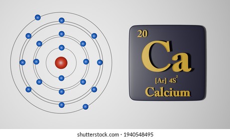
Ca Atoms

Examples of PDB Format
Glucagon is a small protein of 29 amino acids in a single chain.The first residue is the amino-terminal amino acid, histidine,which is followed by a serine residue and then a glutamine.The coordinate information (entry 1gcn) starts with:
Notice that each line or record begins with the record type ATOM.The atom serial number is the next item in each record.
The atom name is the third item in the record.Notice that the first one or two characters of the atom nameconsists of the chemical symbol for the atom type.All the atom names beginning with C are carbon atoms; Nindicates a nitrogen and O indicates oxygen.In amino acid residues, the next character is the remoteness indicator code, which istransliterated according to:
| α | A |
| β | B |
| γ | G |
| δ | D |
| ε | E |
| ζ | Z |
| η | H |

The next data field is the residue type.Notice that each record contains the residue type.In this example, the first residue in the chain is HIS (histidine)and the second residue is a SER (serine).
The next data field contains the chain identifier, in this case A.
The next data field contains the residue sequence number.Notice that as the residue changes from histidine to serine,the residue number changes from 1 to 2.Two like residues may be adjacent to one another,so the residue number is important for distinguishing between them.
The next three data fields contain the X, Y, and Z coordinate values,respectively. The last three fields shown are the occupancy,temperature factor (B-factor), and element symbol.
The spacing of the data fields is crucial.If a data field does not apply, it should be left blank.
The glucagondata file continues in this manner until the final residue is reached:Note that this residue includes the extra oxygen atom OXTon the terminal carboxyl group. Other than OXT and the rarely seen HXT,atoms in standard nucleotides and amino acids in version 3.0 PDBfiles are named according to the IUPAC recommendations (Markley et al.,Pure Appl Chem70:117 (1998)).The TER record terminates the amino acid chain.
A more complicated protein, hemoglobin, consists of four amino acid chains, each with an associated heme group. There are two alpha chains (identifiersA and C) and two beta chains (identifiers B and D).The first ten lines of coordinates for this molecule (entry 3hhb) are:At the end of chain A, the heme group records appear:The last residue in the alpha chain is an ARG (arginine).Again, the extra oxygen atom OXT appears in the terminal carboxyl group.The TER record indicates the end of the peptide chain.It is important to have TER records at the end of peptidechains so a bond is not drawn from theend of one chain to the start of another.
In the example above, the TER record is correct and shouldbe present, but the molecule chain would still be terminated at thatpoint even without a TER record, because HETATM residuesare not connected to other residues or to each other.The heme group is a single residue made up of HETATM records.
After the heme group associated with chain A, chain B begins:
Here the TER card is implicit in the start of a new chain.
Protein Data Bank format relies on the concept of residues:
- Each atom in a residue must be uniquely identifiable.Two atoms in the same residue can only have the same name if theyhave different alternate location identifiers.
- Residue names are a maximum of three characterslong§ and uniquely identify the residue type.Thus, all residues of a given name should be the same type of residueand have the same structure (contain the same atoms with the same connectivity).
Common Errors in PDB Format Files
If a data file fails to display correctly,it is sometimes difficult to determine where in the hundreds of lines ofdata the mistake occurred.This section enumerates some of the most common errors found in PDB files.
Program-Generated PDB Files
Spurious Long Bonds
A couple of common errors in program-generated PDB filesresult in the display of very long bonds between residues:
- Missing TER cards - Either a TER card or a change in the chain IDis needed to mark the end of a chain.
- Improper use of ATOM records instead of HETATM records -HETATM records should be employed for compoundsthat do not form chains, such as water or heme.The first six columns of theATOM record should be changed to HETATM so that theremaining columns stay aligned correctly.
Misaligned Atom Names
Incorrectly aligned atom names in PDB records can cause problems.Atom names are composed of an atomic (element) symbolright-justified in columns 13-14, and trailing identifying charactersleft-justified in columns 15-16. A single-character element symbol should not appear in column 13 unless the atom name has four characters(for example, see Hydrogen Atoms).Many programs simply left-justify all atom names starting in column 13.The difference can be seen clearly in a short segment of hemoglobin(entry 3hhb):
Correct:Incorrect:
Hand-Edited PDB Files
Duplicate Atom Names
One possible editing mistake is the failure to uniquely name all atoms within a given residue.In the following example, two atoms in the same residue are named CA:Depending on the display program, the residue may be shown withincorrect connectivity, or it may become evident only upon labelingthat the residue is missing a CB atom.
Residues Out of Sequence
In the following example, the second residue in thefile is erroneously numbered residue 5.Many display programs will show this residue as connected to residues 1 and 3.If this residue was meant to be connected to residues 4 and 6 instead,it should appear between those residues in the PDB file.
Common Typos
Sometimes the letter l is accidentally substituted for the number 1.This has different repercussions depending on where in the filethe error occurs; a grossly misplaced atom may indicate the presenceof such an error in a coordinate field.These errors can be located readily if the text of the data file appearsin uppercase, by invoking a text editor to search for allinstances of the lowercase letter l.
Hydrogen Atoms
In brief, conventions for hydrogen atoms in version 3.0 PDB formatare as follows:
- Hydrogen atom records follow therecords of all other atoms of a particular residue.
- A hydrogen atom name starts with H. The next part of the nameis based on the name of the connected nonhydrogen atom.For example, in amino acid residues, H isfollowed by the remoteness indicator (if any) of the connected atom,followed by the branch indicator (if any) of the connected atom;if more than one hydrogen is connected to the same atom, an additional digit is appended so that each hydrogen atom will havea unique name. Hydrogen atoms in standard nucleotides and amino acids (other than the rarely seen HXT)are named according to the IUPAC recommendations(Markley et al.,Pure Appl Chem70:117 (1998)).Names of hydrogen atoms in HETATMresidues are determined in a similar fashion.
- If the name of a hydrogen has four characters, it is left-justified starting in column 13; if it has fewer than four characters, it is left-justified starting in column 14.
Ca Atom Electrons
,atom H is attached to atom N. Atom HA is attached to atom CA;the remoteness indicator A is the same for these atoms.Two hydrogen atoms are connected to CB, one is connected to CG,three are connected to CD1, and three are connected to CD2.PQR Variant of PDB Format
Several programs use a modified PDB format called PQR, in whichatomic partial charge (Q) and radius (R) fields follow the X,Y,Z coordinate fields in ATOM and HETATM records. An excerpt:PQR format is rather loosely defined and varies according to whichprogram is producing or using the file. For example, APBSrequires only that all fields be whitespace-delimited.
Ca Atom Charge
If an ATOM or HETATM record being read by Chimera is not in PDB format,Chimera next tries to read it as PQR format. In that case,all fields up to and including the coordinates are still expectedto adhere to the standard format, but the next two eight-column fields are each expected to contain a floating-point number: charge is read fromcolumns 55-62 and radius is read from columns 63-70.The values are assigned as the atom attributescharge and radius, respectively.
PDB2PQRis a program for structure cleanup, charge/radius assignment,and PQR file generation. See also thePDB2PQRtool in Chimera.
Ca Atomic Size
UCSF Computer Graphics Laboratory / May 2020